"The basic structural and functional unit of life".
All living things are composed of cells, almost all of them too
small to see with the naked eye. Although there are exceptions,
a typical eukaryotic cell is 10 to 100 micrometers (μm) (10 to
100 millionths of a meter) in diameter, while most prokaryotic
cells are only 1 to 10 μm in diameter.
The Cell Theory is the Unifying Foundation of Biology
Because cells are so small, they were not discovered until the
invention of the microscope in the 17th century. Robert Hooke was
the first to observe cells in 1665, naming the shapes he saw in cork
cellulae (Latin, “small rooms”). This has come down to us as
cells. Another early microscopist, Anton van Leeuwenhoek , first
observed living cells, which he termed “animalcules,” or little animals. After these early efforts, a century and a half passed before
biologists fully recognized the importance of cells. In 1838, botanist Matthias Schleiden stated that all plants “are aggregates of
fully individualized, independent, separate beings, namely the
cells themselves.” (that all plant tissue was composed of cells). A year later,in 1839, one of his countryman Theodor Schwann reported that all animal tissues also consist of individual cells. He described animal cells as
being similar to plant cells, an understanding that had been long
delayed because animal cells are bounded only by a nearly invisible plasma membrane rather than a distinct cell wall characteristic of plant cells. Schleiden and Schwann are thus credited with the
unifying cell theory that ushered in a new era of productive exploration in cell biology. Another German, Rudolf Virchow, recognized that all cells came from preexisting cells (1858).
Thus, the cell theory was born.
The cell theory developed from the observation that
all organisms are composed of cells. While it sounds simple, it
is a far-reaching statement about the organization of life. In its
modern form, the cell theory includes the following three
principles:
1. All organisms are composed of one or more cells, and the
life processes of metabolism and heredity occur within
these cells.
2.Cells are the smallest living things, the basic units of
organization of all organisms.
3.New cells arises only by division of preexisting cells.
Cells are thought to have evolved spontaneously over 3.5 billion years ago. Biologists have concluded that no cells are originating spontaneously at present. Rather, life on Earth represents a
continuous line of descent from those early cells.
Cell Size is Limited
Most cells are relatively small. Why? The reason relates to the diffusion of substances into and out of cells. The rate of this diffusion
is affected by a number of variables, including (1) the surface area
available for diffusion, (2) temperature, (3) concentration gradient
of the diffusing substance, and (4) distance over which diffusion
must occur. These are related by an equation known as Fick’s Law
of Diffusion. As the size of a cell increases,
the length of time for diffusion from the outside membrane to the
interior of the cell increases as well. This soon becomes a problem, as larger cells need to synthesize more macromolecules, have
correspondingly higher energy requirements, and produce a
greater quantity of waste. Molecules used for energy and biosynthesis must be transported through the membrane. Any metabolic
waste produced must be removed, also passing through the membrane. The rate at which this transport occurs depends on both the
distance to the membrane and the area of membrane available.
The advantage of small cell size is readily apparent in terms
of the surface-area to volume ratio. As a cell’s size increases, its
volume increases much more rapidly than its surface area. For
a spherical cell, the surface area is proportional to the square of
the radius, whereas the volume is proportional to the cube of the
radius. Thus, if the radii of two cells differ by a factor of 10, the
larger cell will have 102, or 100, times the surface area but 103
, or
1000, times the volume of the smaller cell (figure).
Figure:Surface-area to volume ratio. As a cell gets
larger, its volume increases at a
faster rate than its surface area.
If the cell radius increases by
10 times, the surface area
increases by 100 times, but the
volume increases by 1000 times.
The surface area must be large
enough to meet the metabolic
needs of the volume.
The membrane surrounding the cell plays a key role in controlling cell function, because the cell surface provides the only
opportunity for interaction with the environment, as all substances
enter and exit a cell via this surface. Because small cells have more
surface area per unit of volume than large ones, their control over
cell contents is more effective.
Not all cells are small. Some larger cells function quite efficiently because they have structural features that increase surface
area. For example, some cells, such as skeletal muscle cells, have
more than one nucleus, allowing genetic information to be spread
around a large cell. Cells in the nervous system called neurons are
long, slender cells, some extending more than a meter in length.
Although they are long, they are thin, so that any given point
within the cell is close to the plasma membrane. This permits
rapid diffusion between the inside and the outside of the cell. For
the same reason, many cells in your body are shaped flat, like a
thin plate.
Structural features that can dramatically increase a
cell’s surface area are finger-like projections called microvilli.
The cells that line the small intestine are covered with
microvilli.
Microscopy:Microscopes Allow Us to Visualize Cells
Other than egg cells, not many cells are visible to the naked eye
(figure below). Most are less than 50 μm in diameter, far smaller than
the period at the end of this sentence. How do we study cells if
they are too small to see? To visualize cells, we need the aid of
technology.
Figure:The size of cells and their contents. Except for
vertebrate eggs, which can typically be seen with the unaided eye,
most cells are microscopic in size. Prokaryotic cells are generally
1 to 10 μm across.
1 m = 102 cm = 103 mm = 106 μm = 109 nm
Light Microscope
One way to overcome the limitations of our eyes is to increase
magnification so that small objects appear larger. Modern
light microscopes, which operate with visible light, use two magnifying lenses (and a variety of correcting lenses) to achieve very
high magnification and clarity (table 4.1). The first lens focuses
the image of the object on the second lens, which magnifies it
again and focuses it on the back of the eye. Microscopes that
magnify in stages using several lenses are called compound
microscopes.
Electron Microscope
Light microscopes, even compound ones, are not powerful
enough to resolve many of the structures within cells. Why
not just add another magnifying stage to the microscope to
increase its resolving power? The reason we can’t is the limited
resolution of the human eye. Resolution is the minimum distance two points can be apart and still be distinguished as two
separate points. When two objects are closer together than about
100 μm, the light reflected from each strikes the same photoreceptor cell at the rear of the eye. Only when the objects are farther than 100 μm apart can the light from each strike different
cells, allowing your eye to resolve them as two distinct objects
rather than one.Making matters worse, when light beams reflecting from
the two images are closer than a few hundred micrometers, they
start to overlap each other. The only way two light beams can get
closer together and still be resolved is if their wavelengths are
shorter. One way to avoid overlap is to use a beam of electrons
rather than a beam of light. Electrons have a much shorter wavelength, and an electron microscope, employing electron beams,
has 1000 times the resolving power of a light microscope. In
transmission electron microscopes the electrons used to visualize
the specimens are transmitted through the material and are capable of resolving objects only 0.2 nm apart—which is only twice the
diameter of a hydrogen atom!
A second kind of electron microscope, the scanning electron
microscope, bounces beams of electrons off the surface of the specimen. The electrons reflected back, and others that the specimen itself
emits, are amplified and transmitted to a screen, where the image can
be viewed and photographed as a striking three-dimensional image.
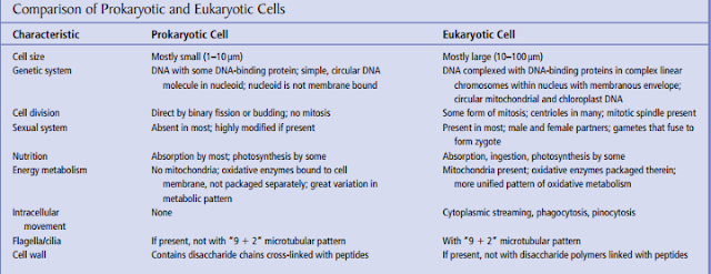
Prokaryotic and Eukaryotic Cells
A fundamental distinction, expressed
in their names, is that prokaryotes lack the membrane-bound
nucleus present in all eukaryotic cells. Among other differences,
eukaryotic cells have many membranous organelles (specialized structures that perform particular functions within cells)
(Table below)
Despite these differences, which are of paramount importance in cell studies, prokaryotes and eukaryotes have much in
common. Both have DNA, use the same genetic code, and synthesize proteins. Many specific molecules such as ATP perform
similar roles in both. These fundamental similarities imply common ancestry. The upcoming discussion is restricted to eukaryotic cells, of which all animals are composed.


